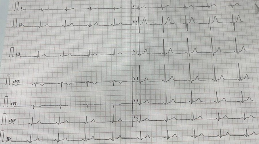
History: A 55 yr old male presented to ED with severe left-sided chest pain from 20 minutes radiating to the left arm associated with SOB and diaphoresis. Bp – 140/82mmhg, P – 84 bpm, spo2 – 92% on RA. ECG done. What’s the diagnosis?
Answer: Posterior MI is suggested by the following changes in V1-3:
- Horizontal ST depression
- Tall, broad R waves (>30ms)
- Upright T waves
- Dominant R wave (R/S ratio > 1) in V2
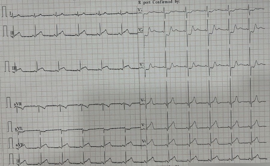
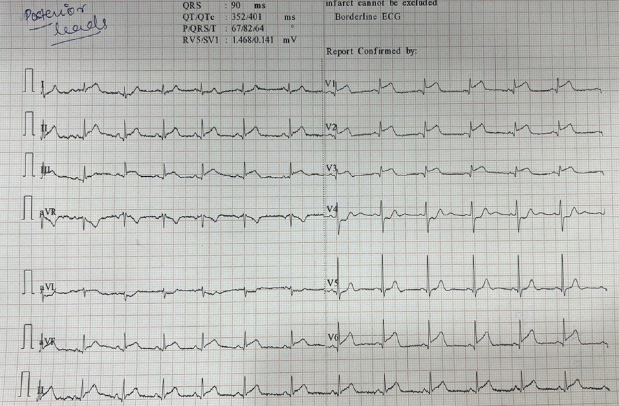
Posterior infarction is confirmed by the presence of ST elevation and Q waves in the posterior leads (V7-9). Leads V7-9 are placed on the posterior chest wall in the following positions (see diagram below):

Source: LITFL
OR you can invert the ECG to see a typical STEMI. For example:

Source: LITFL
The above patient was taken for thrombolysis after stabilization. ECG after thrombolysis.