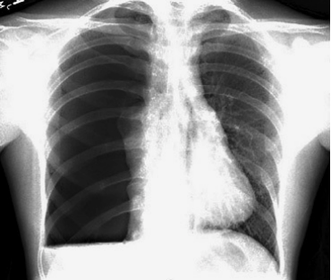
History: A 19-year-old male presents with a rapid onset of shortness of breath. There is no history of trauma or chronic medical disease. The patient has no risk factors for thromboembolic disease. The following x-ray is consistent with which of the following conditions?
- Atypical pneumonia
- Spontaneous pneumothorax
- Metabolic bone disease
- Hampton hump
Anwer: The x-ray demonstrates a spontaneous pneumothorax on the right with a small hemothorax in an otherwise healthy young individual. Management of primary pneumothorax includes aspiration of pleural air either by needle thoracostomy or small catheter with or without Heimlich valve. A Hampton hump is a classic sign of pulmonary embolism with pulmonary infarction that is described as a pleural-based triangular wedge with base along the pleural surface and the top of the triangle pointing toward hilum. This is opposed to Westermark sign that is a sign of vascular oligemia distal to the location of a pulmonary embolism and is described as dilation of proximal pulmonary arteries and collapse of distal vessels. An atypical pneumonia would have a different radiographic appearance including patchy or subsegmental infiltrate. There is no evidence of cortical irregularities or displaced rib fractures on this chest radiograph and there is no history of trauma. Metabolic bone disease would likely manifest with some bony lesion either as lytic lesions or osteopenia.