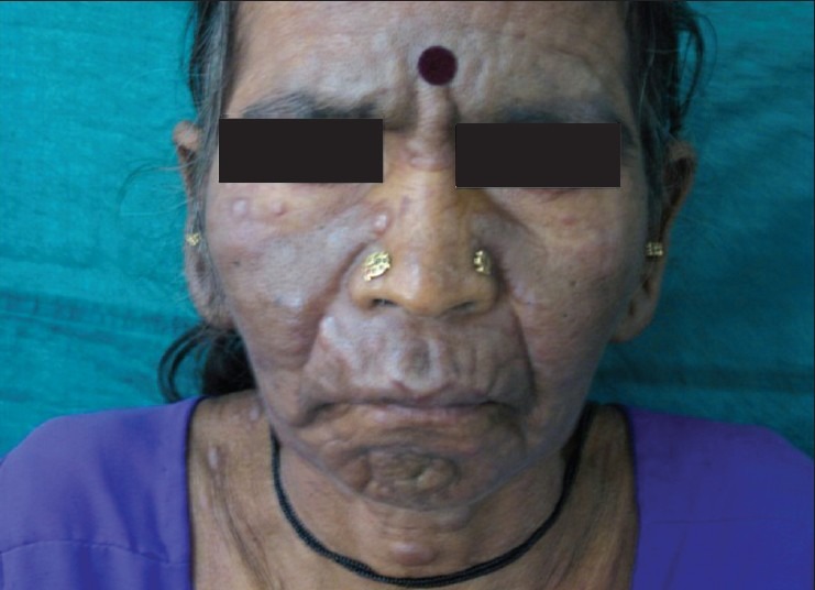
History: A 50-year-old woman presented with asymptomatic multiple cutaneous nodules all over the body of 4 months duration. Cutaneous examination showed multiple hyperpigmented nodules and plaques involving face, trunk, and extremities. Peripheral smear showed abnormally elevated leucocyte count (TLC-70,000) with abnormal cells: myeloblasts 40%, promyelocytes 8% and myelocytes 39%. Auer rods were present in few myeloblasts. What is the most likely diagnosis?
- Gottron’s papules
- Guttate psoriasis
- Immune thrombocytopenia purpura (ITP)
- Leukemia cutis
- Solar purpura
Answer:
Leukemia cutis is the infiltration of neoplastic leukocytes or their precursors into the epidermis, the dermis, or the subcutis, resulting in clinically identifiable cutaneous lesions. Leukemia cutis may follow, precede or occur concomitantly with the diagnosis of systemic leukemia.
The clinical appearance of leukemia cutis is variable with erythematous to violaceous papules or nodules being the most frequent lesions (60%), followed by infiltrated plaques, generalised cutaneous eruption and erythroderma. They are frequently asymptomatic. The nodules are typically firm or rubbery in consistency, and can become purpuric if the patient is thrombocytopenic.