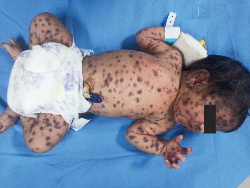
History: A female infant delivered at term had a “blueberry muffin” rash at birth. Laboratory tests and imaging studies were normal. Skin biopsy showed a dense infiltrate of cells with kidney-shaped nuclei and positive S100+ and CD1a+ on immunohistochemistry. What is the most likely diagnosis?
Answer:
Langerhans cell histiocytosis (LCH) is an abnormal clonal proliferation of Langerhans cells, abnormal cells deriving from bone marrow and capable of migrating from the skin to lymph nodes.
Symptoms range from isolated bone lesions to multisystem disease. LCH is part of a group of syndromes called histiocytoses, which are characterized by an abnormal proliferation of histiocytes.
The disease has gone by several names, including Hand–Schüller–Christian disease, Abt-Letterer-Siwe disease, Hashimoto-Pritzker disease (a very rare self-limiting variant seen at birth) and histiocytosis X, until it was renamed in 1985 by the Histiocyte Society.
Diagnosis: Assessment of endocrine function and bone marrow biopsy is also performed when indicated. S-100 protein, CD1a, langerin (CD207).
Treatment: Radiation therapy or chemotherapy