Category

Medical Cases

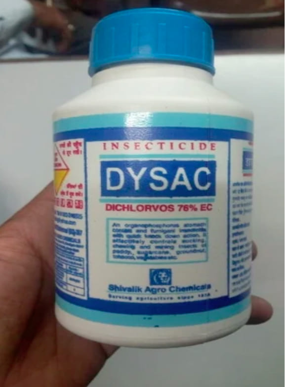
Dichlorvos Ingestion – An OP poisoning case

History: A 27 yr old female brought to ED with A/H/O ingestion of 50ml of Dichlorvos solution at her home 35 minutes ago. On arrival primary survey was

A – Airway compromised

B – Breathing Laboured, Spo2 – 37% on RA, B/L crepts +nt

C – BP – 166/78 mmhg P – 90 bpm

D – E1V2M2 Pupils – B/L constricted, RBS – 112 mg/dl

ABG done shows ph 6.98 pco2 23 pO2 68 hco3 13 Lac 17 AG 22. What’s the diagnosis and how will you manage this patient?

Answer: Dichlorvos is an organophosphate compound.

Look for signs & symptoms of OP poisoning & ask the attendants to bring the sample  :

  • Muscarinic manifestations : DUMBELSDiarrhoea; Urinary incontinence; Miosis; Bradycardia, Bronchorrhea and Bronchospasm; Emesis; Lacrimation, Low blood pressure; Salivation and Sweating. Others include abdominal cramps, hypertension, fascicu­lations, muscle weakness, confusion, coma, seizures.
  • Nicotinic manifestations: Twitching, fasciculations, weakness, diminished respiratory effort, HTN & tachycardia.
  • CNS manifestations: Anxiety, restlessness, tremors, convulsions, confusion, weakness and coma.

Diagnosis:

  • Clinical diagnosis is based on the toxidrome
  • History of exposure
  • ABG, ECG (Prolong QT), CBC, LFT, KFT
  • Cholinesterase assay if available

Rx plan: 

  1. Evaluate ABCD.
  2. Stable patient: Decontamination (Healthcare workers should wear PPE).
  3. Unstable patient: Resuscitation first followed by decontamination. (Never use succinylcholine for intubation).
  4. Gastric lavage within first 1hr of ingestion (Seal GL sample for MLC) with NS & activated charcoal is given orally at a dose of 1-2 g/kg body weight.
  5. Inj. Atropine 1mg or 0.01 to 0.04 mg/kg IV. Double the dose every time and target end points is atropinisation (Clear chest on auscultation with no wheeze, HR>80, pupils no longer pinpoint, dry axilla & SBP >90 mmHg). Once atropinised set up an infusion of atropine at an hourly dose of 10-20% of the total dose of atropine given initially.
  6. Inj. Pralidoxime (2-PAM) is a cholinesterase reactivator. It is effective for nicotinic as well as muscarinic features of toxicity. 30 mg/kg initially over 20 mins followed by a constant infusion at 9 mg/kg/ hour. It is usually continued for 12-24 hrs.

Complications: 

  • Intermediate Syndrome – Syndrome of muscular paralysis occurs within 24-96 hours after ingestion of an organophosphate and following treat­ment of acute cholinergic syndrome. Muscle weakness affects predominantly neck flexors, proximal limb muscles, those supplied by cranial nerves & respiratory muscles.
  • Organophosphate-lnduced Delayed Polyneuropathy (OPIDN) – Cramping muscle pain in the lower limbs, distal numbness and paraesthesia followed by progressive weakness, depres­sion of deep tendon reflexes in the lower limbs > the upper limbs.

Ddx :

  • Carbamate toxicity
  • Nicotine toxicity
  • Methacholine toxicity
  • Arecoline toxicity
  • Bethanechol toxicity
  • Pilocarpine toxicity
  • Pyridostigmine toxicity
  • Neostigmine toxicity
  • Mushroom poisoning
  • Poison hemlock
Clinical Rounds by ClinicHours

Raccoon eyes case

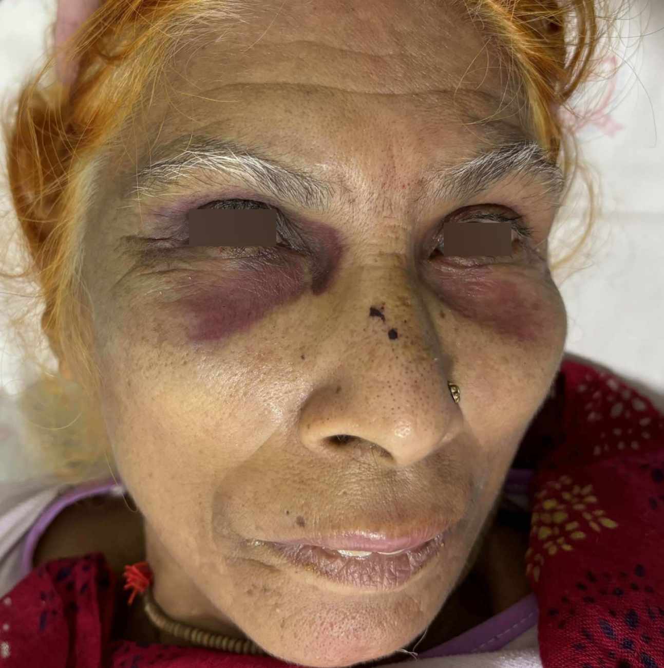
History: A 60 yr female with history of fall from 5 stairs at her residence. No history of LOC/ ENT bleed/ seizure/ vomiting/ FND. On examination B/L periorbital discoloration can be noted. What’s the likely diagnosis?

Answer: Raccoon eyes sign aka panda eyes is periorbital ecchymosis with sparing of the tarsal plate and is a physical examination finding indicative of a base of skull fracture of the anterior cranial fossa. It has positive predictive value of 85%. Also seen in metastatic neuroblastoma, Kaposi sarcoma, multiple myeloma, and amyloidosis.

Clinical Rounds by ClinicHours

A Hydrofluoric acid burn case

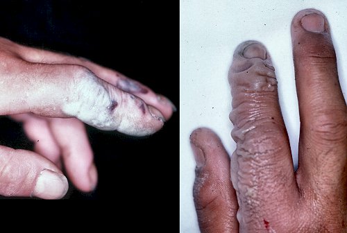
History: A 35 yr old male working with germicides accidentally came in contact with hydrofluoric acid presented to ED with blue-gray appearance with surrounding erythema of skin with extreme pain. How will you manage this in your ED?

Answer:  Decontaminate the patient by removing contaminated clothing and do copious irrigation for 15-30 mins.

  • Apply 2.5 % Calcium gluconate gel LA.
  • Inj 10% Calcium gluconate ID at rate of 0.5mL/cm2 OR 40 ml D5 + 10 mL 10%  calcium gluconate infusion over 4 hrs.

Investigation: VBG, ECG, serum electrolytes.

Hydrofluoric acid (HF) quickly penetrates the skin, where fluoride ions react with calcium and magnesium, causing significant electrolyte disturbances such as hypocalcemia and hypomagnesemia. Additionally, HF inhibits potassium channels, leading to hyperkalemia. These abnormalities can trigger severe cardiac arrhythmias, including ventricular fibrillation, which can be fatal. A notable symptom of HF burns is intense pain disproportionate to the injury, attributed to potassium depletion from nerve endings.

Clinical Rounds by ClinicHours

Posterior wall myocardial Infarction case

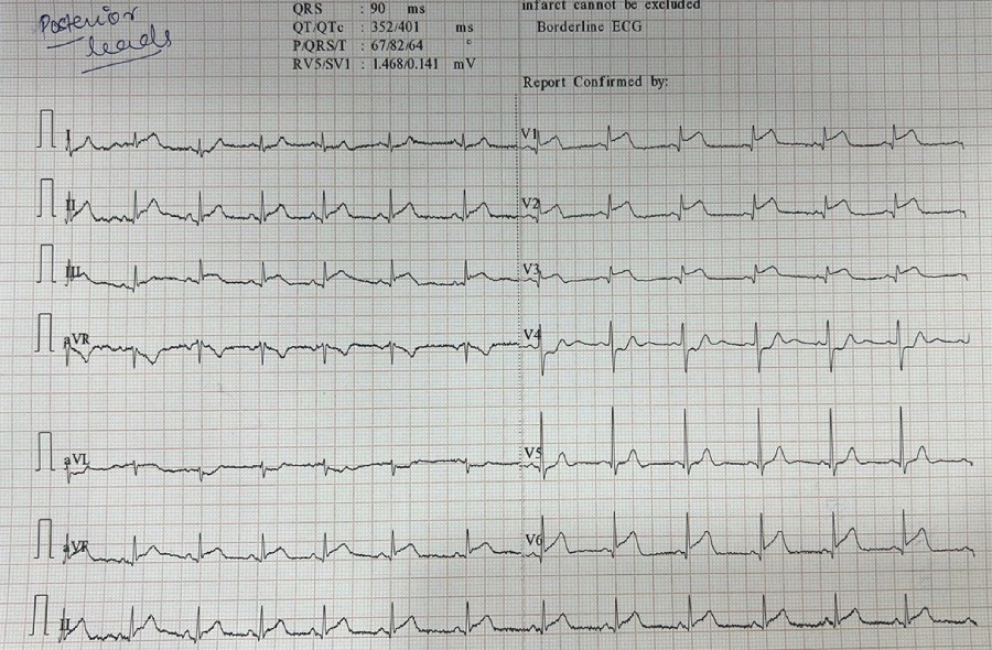
History: A 55 yr old male presented to ED with severe left-sided chest pain from 20 minutes radiating to the left arm associated with SOB and diaphoresis. Bp – 140/82mmhg, P – 84 bpm, spo2 – 92% on RA. ECG done. What’s the diagnosis?

Answer: Posterior MI is suggested by the following changes in V1-3:

  • Horizontal ST depression
  • Tall, broad R waves (>30ms)
  • Upright T waves
  • Dominant R wave (R/S ratio > 1) in V2

Posterior infarction is confirmed by the presence of ST elevation and Q waves in the posterior leads (V7-9). Leads V7-9 are placed on the posterior chest wall in the following positions (see diagram below):


Posterior leads V7 V8 V9 ECG placement

Source: LITFL

OR you can invert the ECG to see a typical STEMI. For example:

ECG Posterior AMI flip image V2

Source: LITFL

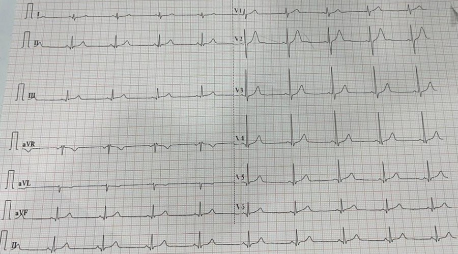
The above patient was taken for thrombolysis after stabilization. ECG after thrombolysis.

Clinical Rounds by ClinicHours

Atrial fibrillation in CKD patient case

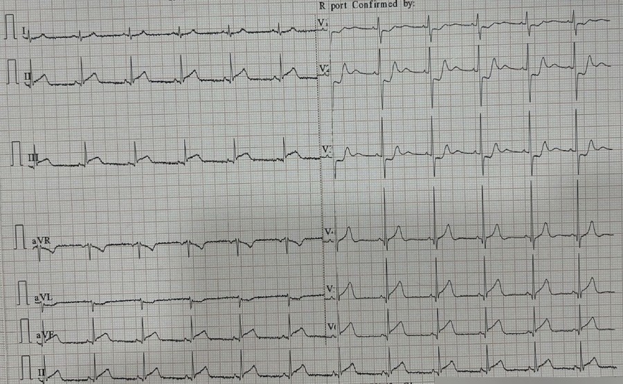
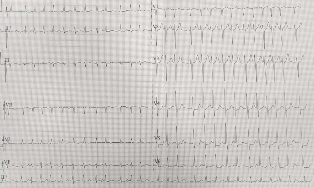
History: A 67-year-old male k/c/o CKD-5 on MHD thrice weekly with Type2DM & HTN presented to ED with SOB, anasarca and AMS. P – 164 bpm irregular, SP02- 80 % on RA, RR – 30, R/E – B/L AE ↓ with B/L crepitations +nt. E3V2M5. ABG – pH 7.17, pCO2 9.7, Na+ 135, k+ 4.5, Lac 10.7, Glu 141, HCO3- 3.6. ECG was done. What’s the diagnosis?

Answer: ECG shows irregularly irregular narrow complex tachycardia with absent P waves & ST ↓ V4-V6 which is s/o Atrial fibrillation.

Classification is dependent on the presentation and duration of atrial fibrillation as below:

First episode – initial detection of AF regardless of symptoms or duration
Recurrent AF – More than 2 episodes of AF
Paroxysmal AF – Self-terminating episode < 7 days
Persistent AF – Not self-terminating, duration > 7 days
Long-standing persistent AF – > 1 year
Permanent AF – Duration > 1 yr in which rhythm control interventions are not pursued or are unsuccessful.

How will you manage the above patient in the ED? Write down in the comment section. 

Clinical Rounds by ClinicHours

Intracranial hemorrhage in CKD patient case

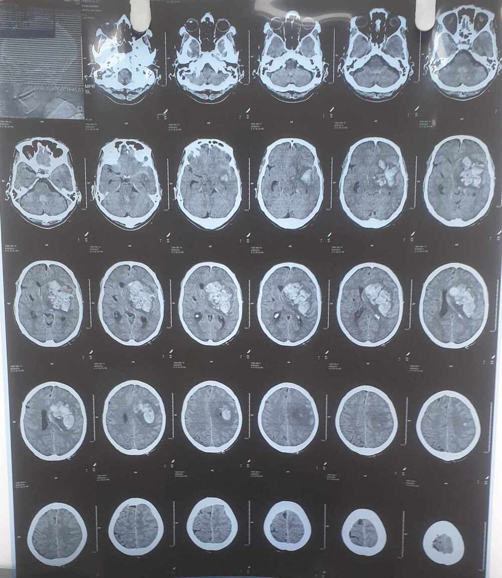
History: A 47-year-old male was brought to ED in an unresponsive state with h/o 1 episode of seizure at home. He is k/c/o CKD5 on MHD twice weekly. BP – 200/120 mmHg, P -118 bpm, Sp02 – 92 % on RA. GCS – E2V1M4. B\L plantar – mute. RBS – 124 mg/dl. ABG (on o2 support) – PH 7.35 PCO2 34.4 PO2 106  HCO3 19.3 Lac – 4. NCCT head was done. What’s the diagnosis?

Answer: NCCT head reveals Intraparenchymal hemorrhage bleed with surrounding edema in left fronto-parieto-temporal, left gangliocapsular region and thalamus extending to the ventricular region with midline shift towards the contralateral side. The patient was intubated i/v/o low GCS and was admitted to the neurosurgery ICU after giving Inj. Levetiracetam 1.5 gm, Inj. Furosemide 40mg, IVF 3% Nacl.

Clinical Rounds by ClinicHours

Port wine stain case

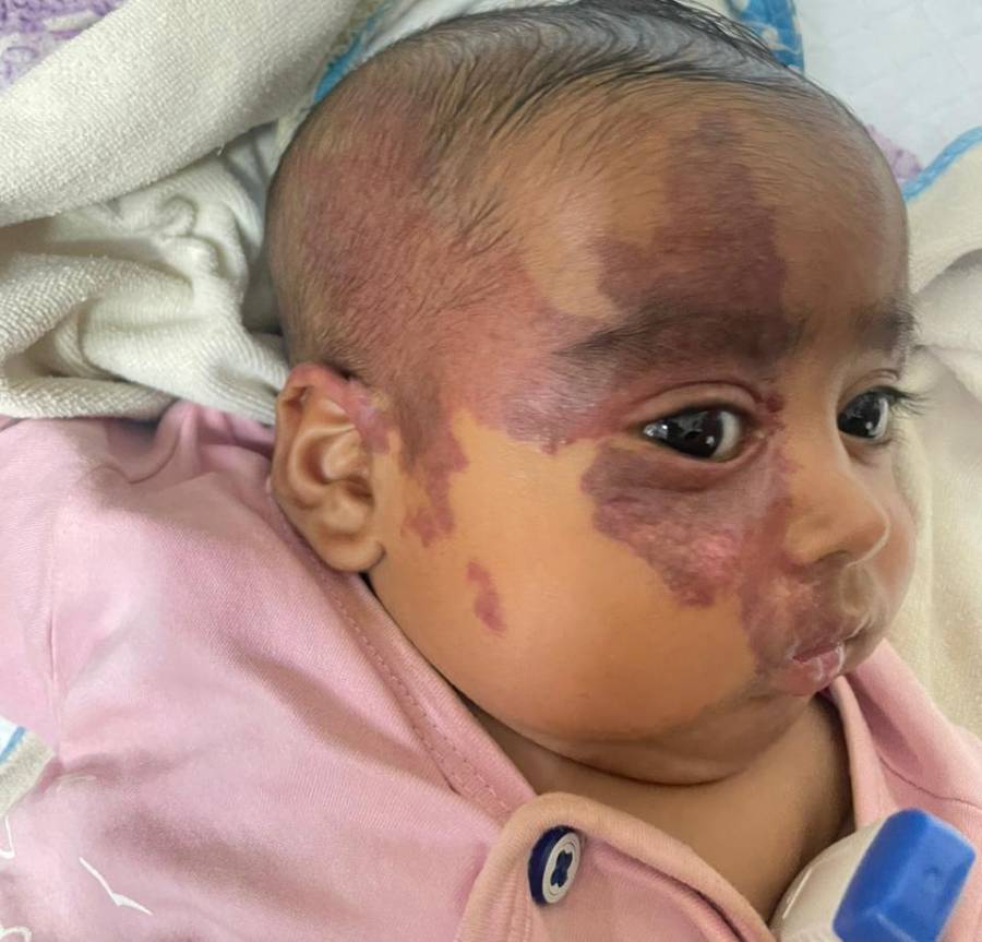
History: A 8-month girl was brought to Dermatology OPD by her mother with a diffuse reddish macular lesion on the right side of her face present since birth. Lesion was smaller, lighter in colour & gradually increased in size. What’s the diagnosis?

Intro: Port wine stain (nevus flammeus) is a discoloration caused by a capillary malformation in the skin. Named after fortified red wine from Portugal.

Cause: Mutation in GNAQ gene on chromosome 9q21 & RASA1 gene. Associated with Sturge-Weber syndrome or Klippel–Trénaunay-Weber syndrome.

Types: Nevus flammeus nuchae, Midline nevus flammeus.

CF: Flat & pink asymptomatic macular patch usually seen at birth or may be acquired. Color may deepen to a dark red or purplish color in adults. Common sites are face, head, neck, abdomen, legs, or arms.

Dx: History based, skin biopsy, MRI Brain (R/O Sturge–Weber syndrome).

Rx:  Pulse Dye or Nd: YAG or KTP Laser, surgery excision, radiation, tattooing, rapamycin LA. If left untreated, these vascular lesions may deepen in color or may undergo hypertrophy & nodular thickening.

Clinical Rounds by ClinicHours

Foreign body Ingestion case

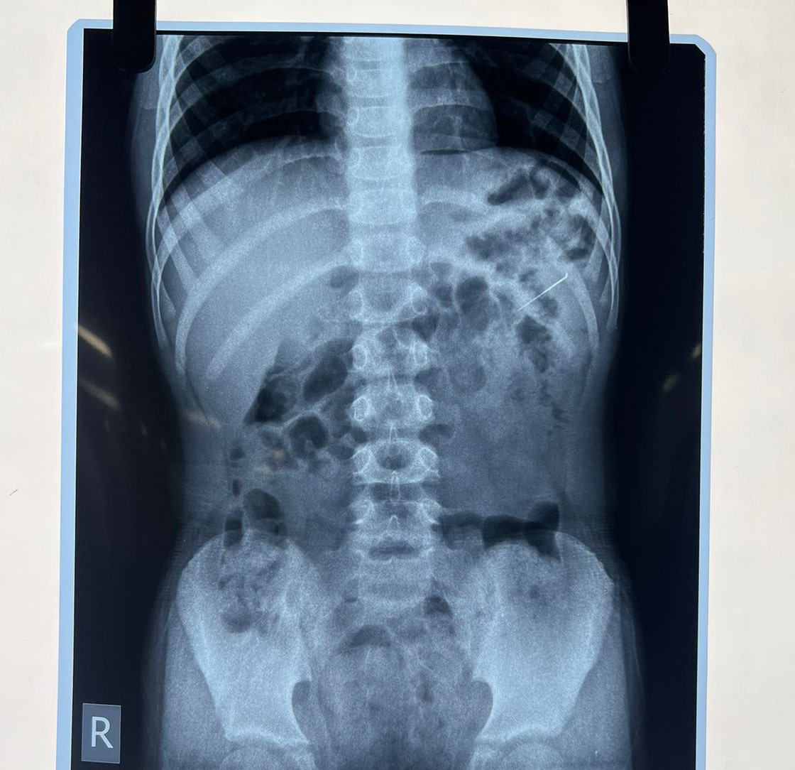
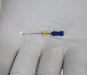
History: A 8 yr old boy came to the ED with accidental ingestion of an Endodontic file while having a dental procedure. Pt. was asymptomatic at the time of presentation with no SOB, abdominal complaints & vomiting. What’s the next step you will do in ED?

Answer: CXR -AP view & abdominal x-ray – Erect & supine was done on arrival after initial assessment. Pt. was hemodynamically stable. Pt was admitted to the surgery ward for observation. There were no signs of peritonitis or perforation. In this case, FB passed spontaneously with the stool on its own & no surgical intervention was required.

Foreign body ingestion is a common clinical problem. Commonly seen in children, older people, people with intellectual disability, psychiatric pathologies & prisoners/inmates.

Most ingested foreign bodies will pass through GI tract without symptoms & cause only minor mucosal injury. However, 10% – 20% of cases will require non-operative intervention, 1% may develop complications (e.g. bowel obstruction, perforation, severe hemorrhage, abscess formation, or septicemia) & require surgical interventions.

Look for dysphagia, abdominal pain, signs of peritonitis, stridor, wheezing, gagging, nausea/vomiting, neck/throat pain, atypical chest pain or non-cardiac chest pain, choking & LGIB.

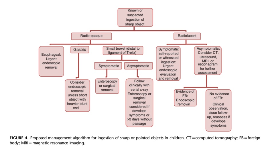
Endoscopy is the first-line intervention for the removal of FB. However, Button battery ingestion can be potentially fatal and thus requires immediate intervention. Check out the management algorithm for sharp FB ingestion.

Clinical Rounds by Clinic Hours

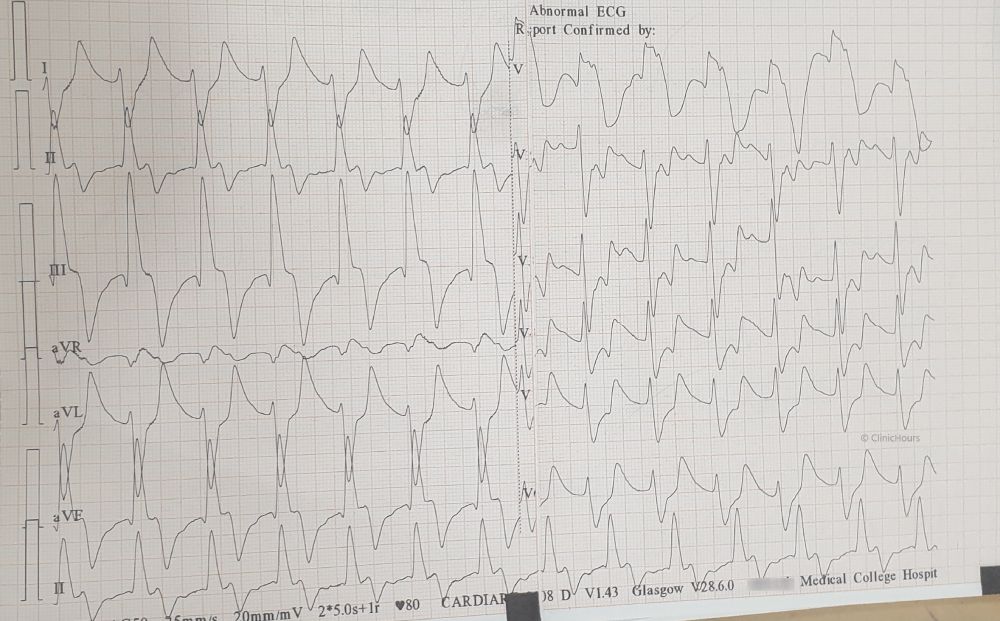
Sine wave appearance in Hyperkalaemia

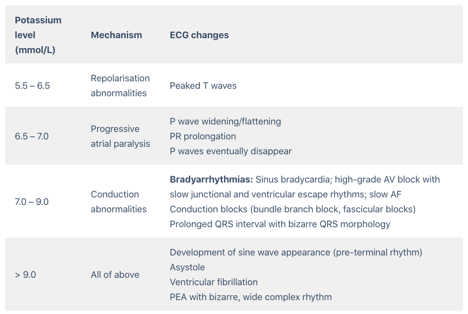
History: A 56 yr old k/c/o CKD-5 on MHD thrice weekly with HTN presented to ED with SOB, drowsy, anasarca. He has missed his hemodialysis this week. BP-170/102 mmhg, P-84 bpm, Spo2 – 82% on RA (99% on NRBM 14/L O2), B\L crepts +nt. Serum K+ – 8.4 mmol/L. What’s the finding in ECG?

Answer: ECG shows sine wave appearance in severe hyperkalemia. Hyperkalaemia is defined as a serum potassium level of > 5.2 mmol/L. ECG changes generally do not manifest until there is a moderate degree of hyperkalaemia (≥ 6.0 mmol/L). The earliest manifestation of hyperkalaemia is an increase in T wave amplitude.

The patient was treated with 10% 10ml of IV calcium gluconate, nebulised with Salbutamol, 10 units of regular insulin IV combined with  dextrose 50%, IV sodium bicarbonate 50 mEq and Inj. Furosemide 40mg IV. Emergent haemodialysis was initiated. Pt was shifted to ICU.

Clinical Rounds by Clinic Hours

Rice grain calcification in cysticercosis

History: A 56 year old female presented to ED with 1 episode of GTC seizure. Neurological examination was normal except for postictal confusion. NCCT brain showed multiple calcified granuloma, some showing perilesional oedema in the frontal, parietal & occipital lobes. Routine CXR showed typical rice-grain-shaped calcification in the chest wall muscles. Xray thigh and forearm were ordered which showed similar lesions. What’s the diagnosis?

Answer: Rice grain calcification is characteristic of infection with Taenia solium (cysticercosis); when the inflammatory response of the host kills the larval cysts (cysticerci), they undergo granulomatous change and become calcified. Demonstration of rice-grain clacification on plain radiograph is a minor diagnostic criterion for neurocysticercosis (NCC). Their presence support NCC as the cause of ring-enhancing lesions in brain imaging.

Clinical Rounds by ClinicHours
error: